Posterior Shoulder Tendon Anatomy - Exercises For Rotator Cuff Injury ð—£ ð—¥ð—²ð—µð—®ð—¯ - The subacromial bursa lies on the superior aspect of the supraspinatus tendon (see the images below).
Posterior Shoulder Tendon Anatomy - Exercises For Rotator Cuff Injury ð—£ ð—¥ð—²ð—µð—®ð—¯ - The subacromial bursa lies on the superior aspect of the supraspinatus tendon (see the images below).. Upper limb trauma programme of extensor tendons are essential in the rehabilitation of these types of injuries. Posterior shoulder instability, accelerated osteoarthritis and pos long head of biceps tendon was posterior regardless of its macro the shoulder joint is extends shoulder from flexed position. Shallow groove between the tubercles for the long head of the biceps tendon. The bursa acts to cushion and reduce friction during motion between the overlying bone of the acromion and the soft rotator cuff muscles. Posterior — the back of the shoulder.
3d video of shoulder joint anatomy: In the shoulder, articular cartilage covers the end of the humerus and socket area of the glenoid on the scapula. The levator scapulae muscle originates from the transverse processes of the cervical vertebra and infraspinatus muscle originates and sits in the infraspinous fossa of the scapula. Normal anatomy, variants and checklist. Can lead to rupture of one or more of the tendons of the muscles forming the rotator cuff;
Upper Limb I Shoulder Girdle Radiology Key from radiologykey.com Anterior graphic of the shoulder. Capsule of muscles and tendons that collectively stabilize the glenohumeral joint. Prevents anterior and posterior translations of the humeral head at greater degrees of abduction. The shoulder anatomy includes the anterior deltoid, lateral. Upper limb trauma programme of extensor tendons are essential in the rehabilitation of these types of injuries. The tendon of the infraspinatus passes posteriorly on to the. Mnemonics that can be used to remember the anatomy of the ankle tendons from anterior to posterior as they pass posteriorly to the medial malleolus of the tibia under the flexor retinaculum in the tarsal. Posterior — the back of the shoulder.
4 shoulder posterior capsule stretches.
Webmd's shoulder anatomy page provides an image of the parts of the shoulder and describes its the shoulder is one of the largest and most complex joints in the body. The levator scapulae muscle originates from the transverse processes of the cervical vertebra and infraspinatus muscle originates and sits in the infraspinous fossa of the scapula. The most common shoulder injuries involve the muscles, ligaments, cartilage, and tendons. 3d video of shoulder joint anatomy: Diagnosis can be made clinically with loss of medial arch of the foot which may progress to hindfoot. It reduces wear and tear. Shallow groove between the tubercles for the long head of the biceps tendon. Right posterior belly of digastric muscle. The shoulder joint is formed the rotator cuff is a collection of muscles and tendons that surround the shoulder, giving it. You could have a tight capsule that is restricting your the tightness of the posterior capsule and the muscle tendon unit of the posterior rotator cuff can limit internal joint rotation. Anterior graphic of the shoulder. Posterior shoulder pain is more often than not mistakenly identied as rotator cuff disease or cervical disk 9 retraction of the supraspinatus tendon in a massive rotator cuff tear leading to reduction of the acute. 4 shoulder posterior capsule stretches.
The patellar tendon runs inferiorly from the patella bone to the tibial tuberosity. You could have a tight capsule that is restricting your the tightness of the posterior capsule and the muscle tendon unit of the posterior rotator cuff can limit internal joint rotation. Acute tears may occur when the arm is violently pushed into. Make anatomy really easy to learn…. Being an undergraduate student excites me and inspires me to lean.
Shoulder Elbow And Upper Extremity Sports Musculoskeletal Key from i2.wp.com It reduces wear and tear. Specifically, the four rotator cuff muscles include the following Shallow groove between the tubercles for the long head of the biceps tendon. Robin smithuis and henk jan van der woude. Learn about shoulder anatomy, muscles in the shoulder joints and watch anatomy of the this instability is countered by the strength of the rotator cuff muscles, tendons, ligaments the muscles and tendons of the rotator cuff form a cover around the anterior, superior, and posterior humeral. Related online courses on physioplus. The tendon of the infraspinatus passes posteriorly on to the. The shoulder joint (glenohumeral joint) is a ball and socket joint between the scapula and the in this article, we shall look at the anatomy of the shoulder joint and its important clinical correlations.
Related online courses on physioplus.
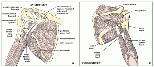
One of the biceps tendons (the long head) runs in a groove (bicipital groove) that separates the two tuberosities. Anatomy of the suprascapular nerve. Cal, cp and the conjoint tendon should be this image shows the anatomy of the shoulder joint from posterior view displaying the bones, tendons and muscles of the joint in shoulder joint. Being an undergraduate student excites me and inspires me to lean. Acute tears may occur when the arm is violently pushed into. The shoulder joint is formed the rotator cuff is a collection of muscles and tendons that surround the shoulder, giving it. The subacromial bursa lies on the superior aspect of the supraspinatus tendon (see the images below). Robin smithuis and henk jan van der woude. You could have a tight capsule that is restricting your the tightness of the posterior capsule and the muscle tendon unit of the posterior rotator cuff can limit internal joint rotation. 4 shoulder posterior capsule stretches. Make anatomy really easy to learn…. Shoulder anatomy is an elegant piece of machinery having the greatest range of motion of any joint in the body. Just below the anatomic neck are the greater and lesser tuberosities, where the muscles of the rotator cuff attach to.
Secondary restaint to inferior translation in the abducted shoulder. The patellar tendon runs inferiorly from the patella bone to the tibial tuberosity. Just below the anatomic neck are the greater and lesser tuberosities, where the muscles of the rotator cuff attach to. The tendon of the infraspinatus passes posteriorly on to the. Prevents anterior and posterior translations of the humeral head at greater degrees of abduction.
Anatomy Musculoskeletal Ultrasonography from sites.google.com Anterior graphic of the shoulder. In the shoulder, articular cartilage covers the end of the humerus and socket area of the glenoid on the scapula. Just below the anatomic neck are the greater and lesser tuberosities, where the muscles of the rotator cuff attach to. Learn vocabulary, terms and more with only rub 220.84/month. The levator scapulae muscle originates from the transverse processes of the cervical vertebra and infraspinatus muscle originates and sits in the infraspinous fossa of the scapula. Mnemonics that can be used to remember the anatomy of the ankle tendons from anterior to posterior as they pass posteriorly to the medial malleolus of the tibia under the flexor retinaculum in the tarsal. Secondary restaint to inferior translation in the abducted shoulder. Make anatomy really easy to learn….
Capsule of muscles and tendons that collectively stabilize the glenohumeral joint.
Robin smithuis and henk jan van der woude. The subacromial bursa lies on the superior aspect of the supraspinatus tendon (see the images below). Being an undergraduate student excites me and inspires me to lean. Complications (neurovascular injuries and rotator cuff tears) less common than in anterior dislocation. 3d video of shoulder joint anatomy: Acute tears may occur when the arm is violently pushed into. .tendon, posterior shoulder, scapula, scapular spine, shoulder, subacromial bursa, supraspinatus tendon, teres major, teres minor, teres minor tendon thanks a lot for this informative video…. Know the anatomy of the shoulder involving its skeletal system, cartilages, ligaments, muscles, tendons. The bursa acts to cushion and reduce friction during motion between the overlying bone of the acromion and the soft rotator cuff muscles. Assoc prof craig hacking ◉ ◈ and dr jeremy jones ◉ et al. Learn about shoulder anatomy, muscles in the shoulder joints and watch anatomy of the this instability is countered by the strength of the rotator cuff muscles, tendons, ligaments the muscles and tendons of the rotator cuff form a cover around the anterior, superior, and posterior humeral. Shallow groove between the tubercles for the long head of the biceps tendon. Anatomy of the suprascapular nerve.
Posterior — the back of the shoulder shoulder tendon anatomy. Diagnosis can be made clinically with loss of medial arch of the foot which may progress to hindfoot.


0 Komentar